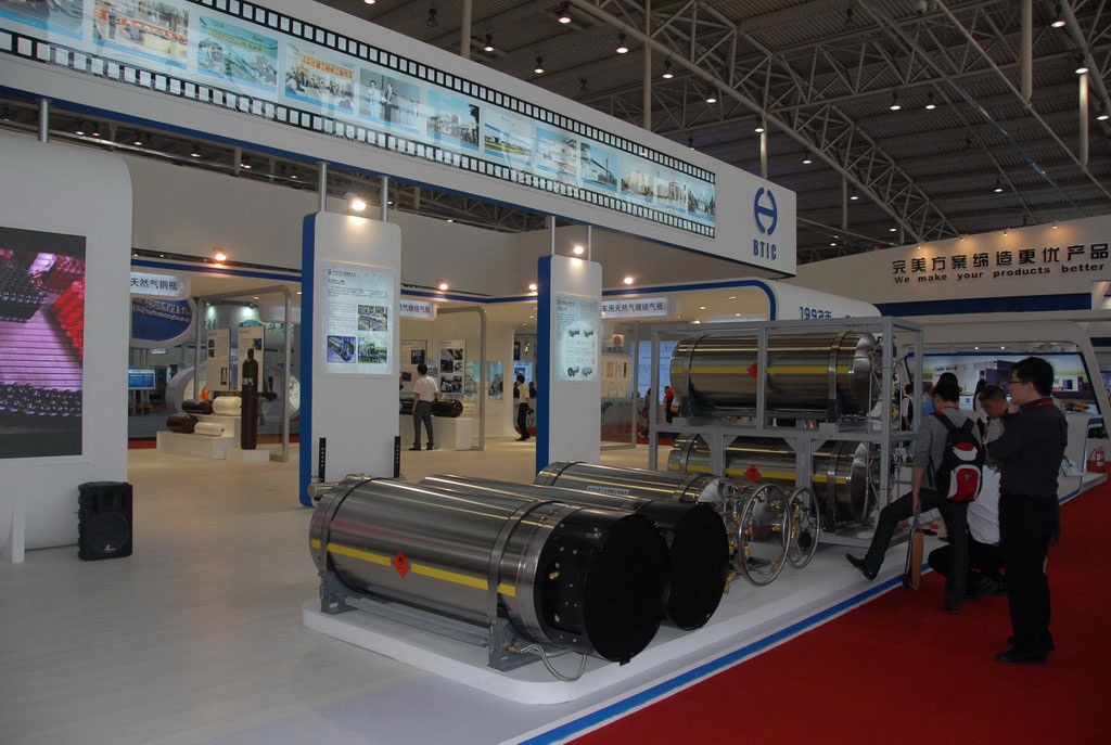
The sugars and phosphates in the backbone of a dna strand are held together by __________.


What do a human, a rose, and a bacterium have in common? Each of these things — along with every other organism on Earth — contains the molecular instructions for life, called deoxyribonucleic acid or DNA. Encoded within this DNA are the directions for traits as diverse as the color of a person's eyes, the scent of a rose, and the way in which bacteria infect a lung cell.

DNA is found in nearly all living cells. However, its exact location within a cell depends on whether that cell possesses a special membrane-bound organelle called a nucleus. Organisms composed of cells that contain nuclei are classified as eukaryotes, whereas organisms composed of cells that lack nuclei are classified as prokaryotes. In eukaryotes, DNA is housed within the nucleus, but in prokaryotes, DNA is located directly within the cellular cytoplasm, as there is no nucleus available.

But what, exactly, is DNA? In short, DNA is a complex molecule that consists of many components, a portion of which are passed from parent organisms to their offspring during the process of reproduction. Although each organism's DNA is unique, all DNA is composed of the same nitrogen-based molecules. So how does DNA differ from organism to organism? It is simply the order in which these smaller molecules are arranged that differs among individuals. In turn, this pattern of arrangement ultimately determines each organism's unique characteristics, thanks to another set of molecules that "read" the pattern and stimulate the chemical and physical processes it calls for.

What components make up DNA?

At the most basic level, all DNA is composed of a series of smaller molecules called nucleotides. In turn, each nucleotide is itself made up of three primary components: a nitrogen-containing region known as a nitrogenous base, a carbon-based sugar molecule called deoxyribose, and a phosphorus-containing region known as a phosphate group attached to the sugar molecule (Figure 1). There are four different DNA nucleotides, each defined by a specific nitrogenous base: adenine (often abbreviated "A" in science writing), thymine (abbreviated "T"), guanine (abbreviated "G"), and cytosine (abbreviated "C") (Figure 2).

Although nucleotides derive their names from the nitrogenous bases they contain, they owe much of their structure and bonding capabilities to their deoxyribose molecule. The central portion of this molecule contains five carbon atoms arranged in the shape of a ring, and each carbon in the ring is referred to by a number followed by the prime symbol ('). Of these carbons, the 5' carbon atom is particularly notable, because it is the site at which the phosphate group is attached to the nucleotide. Appropriately, the area surrounding this carbon atom is known as the 5' end of the nucleotide. Opposite the 5' carbon, on the other side of the deoxyribose ring, is the 3' carbon, which is not attached to a phosphate group. This portion of the nucleotide is typically referred to as the 3' end (Figure 1). When nucleotides join together in a series, they form a structure known as a polynucleotide. At each point of juncture within a polynucleotide, the 5' end of one nucleotide attaches to the 3' end of the adjacent nucleotide through a connection called a phosphodiester bond (Figure 3). It is this alternating sugar-phosphate arrangement that forms the "backbone" of a DNA molecule.

How is the DNA strand organized?

Although DNA is often found as a single-stranded polynucleotide, it assumes its most stable form when double stranded. Double-stranded DNA consists of two polynucleotides that are arranged such that the nitrogenous bases within one polynucleotide are attached to the nitrogenous bases within another polynucleotide by way of special chemical bonds called hydrogen bonds. This base-to-base bonding is not random; rather, each A in one strand always pairs with a T in the other strand, and each C always pairs with a G. The double-stranded DNA that results from this pattern of bonding looks much like a ladder with sugar-phosphate side supports and base-pair rungs.

Note that because the two polynucleotides that make up double-stranded DNA are "upside down" relative to each other, their sugar-phosphate ends are anti-parallel, or arranged in opposite orientations. This means that one strand's sugar-phosphate chain runs in the 5' to 3' direction, whereas the other's runs in the 3' to 5' direction (Figure 4). It's also critical to understand that the specific sequence of A, T, C, and G nucleotides within an organism's DNA is unique to that individual, and it is this sequence that controls not only the operations within a particular cell, but within the organism as a whole.

How is DNA packaged inside cells?

During DNA packaging, long pieces of double-stranded DNA are tightly looped, coiled, and folded so that they fit easily within the cell. Eukaryotes accomplish this feat by wrapping their DNA around special proteins called histones, thereby compacting it enough to fit inside the nucleus (Figure 8). Together, eukaryotic DNA and the histone proteins that hold it together in a coiled form is called chromatin.

How do scientists visualize DNA?

It is impossible for researchers to see double-stranded DNA with the naked eye — unless, that is, they have a large amount of it. Modern laboratory techniques allow scientists to extract DNA from tissue samples, thereby pooling together miniscule amounts of DNA from thousands of individual cells. When this DNA is collected and purified, the result is a whitish, sticky substance that is somewhat translucent.

To actually visualize the double-helical structure of DNA, researchers require special imaging technology, such as the X-ray diffraction used by Rosalind Franklin. However, it is possible to see chromosomes with a standard light microscope, as long as the chromosomes are in their most condensed form. To see chromosomes in this way, scientists must first use a chemical process that attaches the chromosomes to a glass slide and stains or "paints" them. Staining makes the chromosomes easier to see under the microscope. In addition, the banding patterns that appear on individual chromosomes as a result of the staining process are unique to each pair of chromosomes, so they allow researchers to distinguish different chromosomes from one another. Then, after a scientist has visualized all of the chromosomes within a cell and captured images of them, he or she can arrange these images to make a composite picture called a karyotype (Figure 10).

Breaks in the sugar phosphate DNA backbone that can lead to the exposure of single-stranded breaks or the generation of DNA double-strand breaks (DSBs) are activators of the DDR.

From: Liver Pathophysiology, 2017

From The School of Biomedical Sciences Wiki

The sugar phosphate backbone is an important stuctural component of DNA. It consists of 5-carbon deoxyribose sugars and phosphate groups. These sugars are linked together by a phosphodiester bond, between carbon 4 of their chain, and a CH2 group that is attached to a phosphate ion. They are extremely important in the function of DNA.

The sugars and phosphates in the backbone of a dna strand are held together by __________.

Figure 1 Diagram showing the sugar phosphate backbone of DNA, and the nitrogenous bases attached to it, forming a nucleotide [1]

Structure of DNA

DNA is wound into an right-handed double helix. The strands are anti-parellel i.e. one runs 3' to 5', the other run 5' to 3'. This is done by the sugar phosphate backbone twisting around itself in a coil. The purpose of this twisting is to protect the bases inside it, and prevent them from being damaged by the environment. DNA is very stable due to rungs of “ladder” is hydrophobic and phosphate sugar backbone of DNA is negatively charged. These features make DNA can repel water and would not hydrolysed and breakdown by the aqueous environment. One turn of this helix is 34nm long, the diameter of it is 2nm, and there are ten bases attached per turn at 0.34nm.

Differences in RNA and DNA

RNA and DNA are both examples of phosphodiesters and have a very similar structure. However, their sugar phosphate backbone differs slightly. In DNA, the sugar involved is deoxyribose. On on the other hand, the sugar in the backbone of RNA is called ribose. These two sugars only differ by one -OH group being changed to an -H, but provides different capabilities for each molecule[2].

References

  1. ↑ Krebs J.E. Goldstein E.S. Kilpatrick S.T 2011 Lewin's Genes X, 10th Edition, Jones and Bartlett Publishers: London
  2. ↑ Berg J.M. Tymoczko J.L. Stryer L. 2012 Biochemistry (pages 114-115), 7th Edition, WH Freeman and Company: New York